Keratoconus: What It Is and How It’s Treated
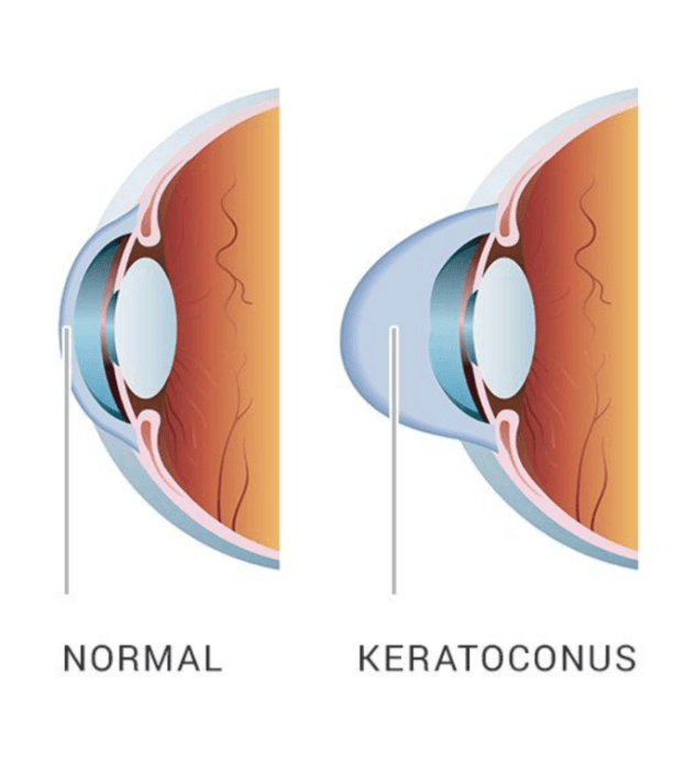 Keratoconus is a progressive eye condition that affects the shape of the cornea - the clear, front surface of the eye. Instead of maintaining a smooth, dome-like shape, the cornea gradually thins and begins to bulge outward into a cone shape.
This change disrupts how light enters the eye, leading to distorted and blurred vision that often cannot be fully corrected with standard glasses.
What Causes Keratoconus?
The exact cause of keratoconus is not fully understood, but it is believed to involve a combination of genetic and environmental factors.
Common contributing factors include:
A family history of keratoconus
Chronic eye rubbing
Underlying conditions such as asthma, allergies or eczema
Structural weakness in the corneal tissue
Keratoconus typically develops during the teenage years or early adulthood and may progress over time if left unmanaged. Read more here: The Genetic and Environmental Factors for Keratoconus - PMC
Symptoms of Keratoconus
In its early stages, keratoconus can be difficult to detect. As the condition progresses, symptoms become more noticeable.
These may include:
Blurred or distorted vision
Increased sensitivity to light (photophobia)
Frequent changes in glasses prescription
Difficulty seeing clearly at night
Ghosting or multiple images (double vision in one eye)
Because symptoms can develop gradually, many people may not realise their vision changes are linked to an underlying condition.
How Is Keratoconus Diagnosed?
Keratoconus is diagnosed through a comprehensive eye examination using advanced imaging technology.
This may include:
Corneal topography to map the shape of the cornea
Measurement of corneal thickness
Assessment of visual acuity and refraction
Early detection is important, as it allows for intervention before significant progression occurs.
How Is Keratoconus Treated?
Treatment depends on the severity and progression of the condition. The goal is to improve vision and, where possible, stabilise the cornea.
Glasses and Contact Lenses
In early stages, glasses or soft contact lenses may provide adequate vision correction. As keratoconus progresses, rigid gas permeable (RGP) or specialty lenses may be required to improve visual clarity.
Corneal Cross-Linking
Corneal cross-linking is a minimally invasive procedure designed to strengthen the cornea and slow or halt progression. It works by using riboflavin (vitamin B2) and UV light to reinforce the collagen fibres within the cornea.
Surgical Options
In more advanced cases, surgical intervention may be considered. This can include corneal implants or, in severe cases, a corneal transplant.
Modern treatment approaches aim to intervene early and preserve the natural cornea wherever possible.
Why Early Treatment Matters
Keratoconus is a progressive condition, meaning it can worsen over time if left untreated.
Early diagnosis and appropriate management can help:
Slow or stop progression
Maintain better visual quality
Reduce the need for more invasive procedures
When to Seek Professional Advice
If you are experiencing ongoing changes in your vision, frequent prescription updates or increasing difficulty seeing clearly, it is important to have your eyes assessed.
A tailored evaluation can determine whether keratoconus is present and what treatment options are most appropriate for your eyes. Are you worried? Come in for an appointment and let us help you before things progress.
| Tags:Keratoconus |